Magnetic Resonance Imaging (MRI) is a type of imaging that uses magnetic fields, radio frequency waves, and computers to create highly detailed images. With MRI, doctors can see your internal organs and other structures without the use of ionizing radiation.
We offer music for patient comfort and have a Wide Bore for a more spacious exam
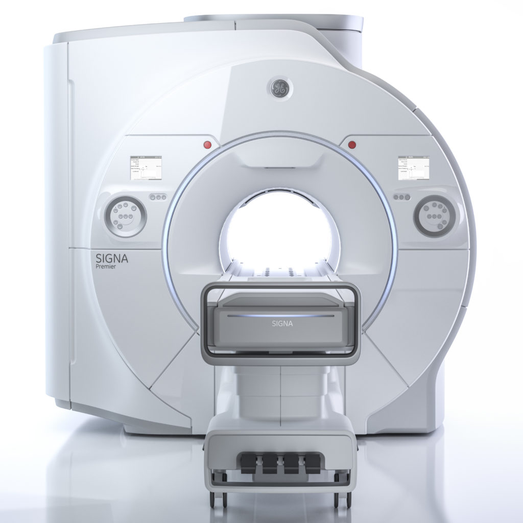
Oaklawn’s MRI system provides the fastest diagnostic magnetic resonance imaging technology available with the utilization of a 3-Tesla Wide Bore. We also offer a variety of music choices through a streaming application.
A 3-tesla magnetic field is twice as powerful as the fields used in conventional high-field MRI scanners, and as much as 15 times stronger than low-field or open MRI scanners. This results in a clearer and more complete diagnostic image. Stronger magnetic fields also produce better, more detailed images of soft tissues and organs than standard, lower strength MRI scanners.
Another advantage of Oaklawn’s 3-Tesla Wide Bore MRI is that the wider machine offers more comfort than typical MRI scanners as some patients may feel anxious or claustrophobic in traditional MRI scanners. A 3-Tesla Wide Bore MRI offers a more spacious bore, or tube, and in some cases the patient’s head can remain outside of the bore for scans that don’t involve the spinal cord, neck, or head. Additionally, most exams can be performed with the patient entering the MRI machine feet first.
How do I schedule an MRI?
- Your primary care provider’s office can schedule you. Choosing CT or MRI Resource Booklet for Ordering Physicians
- You can call scheduling directly at (269) 789-3915, option 2, and make an appointment yourself.
(For the second option you will need to have a copy of your provider’s order with you or your provider’s office will need to fax the order to scheduling at (269) 789-3971)
Where can I have an MRI examination?
200 N. Madison St.
Marshall, MI 49068
MRI Exams Completed at Oaklawn
MRI studies offered at Oaklawn with approximate durations for each test. When contrast is needed for an MRI exam, an additional 10 to 15 minutes may be added to the exam.
Abdomen and Pelvic imaging
- Exam Time: 30 min – 1 hour
- Prep: Refrain from eating or drinking anything for 8 hours prior to testing
Arterial Head, Neck, or Abdomen imaging
- Exam Time: 15 – 30 min
Arthrogram imaging, including an injection by a Radiologist in the Xray Fluoroscopy department
- Exam Time: 1 hour & 15 min
Brain, Face, Neck, and Orbit imaging
- Exam Time: 20 min – 1 hour & 15 min
Breast and Chest imaging
- Exam Time: 30 – 45 min
Extremity imaging such as Ankle, Elbow, Femur, Foot, Forearm, Hand, Hip, Humerus, Knee, Shoulder, Tibia/Fibula, Wrist imaging
- Exam Time: 30 – 45 min
Cervical, Thoracic, and Lumbar Spine Imaging
- Exam Time: 30 – 45 min for each area
TMJ imaging
- Exam Time: 45 min
Please note these are estimates for each type of MRI exam. Many factors can affect the length of a test and may vary for each patient.
MRI Contrast
Multihance (Used for Arterial and Breast Imaging Only)
Eovist (Used for Abdominal imaging only)
Contrast used in MRI imaging, called Gadolinium, is specific to MRI and is a different chemical compound than the contrast used in CT. MRI contrast will not make a patient feel warm, like CT contrast. Alternatively, a patient may feel a cool sensation go up their arm during the injection, due to the contrast being stored at room temperature and body temperature being warmer. Some patients may not feel this temperature difference, or any sensation upon MRI contrast injection, and that is not cause for concern.
MRI Safety and Implants
MRI Pre-Screening Implant Safety Questions
Patients will have a comprehensive evaluation of their personal history regarding implants, devices, objects, and materials attached-to or implanted within their body from procedures or surgeries prior to their MRI examination.
Always consult the MRI Technologist or Radiologist if you have any question or concerns before entering the MR system room. Do not enter the MR system room or MR environment if you have any question or concern regarding any implant, device, or object.
The MR system magnet is ALWAYS on! Certain metallic, electronic, magnetic, or mechanical implants, devices, or objects may be hazardous to you and/or may interfere with the MRI procedure. Therefore, all individuals are required to fill out a new MRI Patient Screening form before entering the MR environment or MR system room.
Patients are required to remove medication patches, pumps, glucose monitors, and all other metallic objects before entering the MR environment or MR system room. Common items include, but are not limited to: hearing aids, medical alert or tether alarms, beeper, cell phone, keys, eye glasses, hair pins, barrettes, wigs, jewelry (including body piercing jewelry), watch, safety pins, paperclips, money clips, credit cards, bank cards, magnetic strip cards, strip cards, coins, pens, pocket knife, nail clipper, steel-toed boots/shoes, tools, clothing with metal fasteners, and clothing with metallic threads or moisture wicking fabric. Loose metallic objects are especially prohibited in the MR system room and MR environment.
Components within the MRI system vibrate and create loud noises, since the scanner has a hollow inside these sounds echo and can become louder. You will be required to wear facility provided earplugs or hearing protection during the MRI procedure to prevent possible problems related to acoustic noise.